
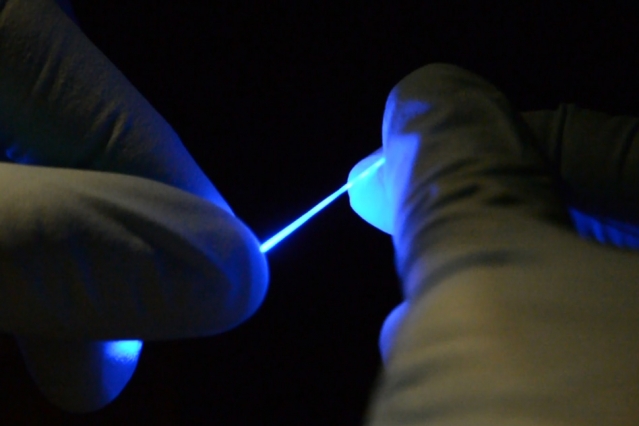
A Center for Sensorimotor Neural Engineering (CSNE)-affiliated team of faculty and student researchers from the Massachusetts Institute of Technology (MIT) and the University of Washington (UW) recently made a significant advance in the design of implantable fibers for studying neurons in the spinal cord. The team developed a flexible and stretchable fiber that can simultaneously deliver optical (light) pulses for neural stimulation and electrical connections for neural recording, as described in their paper released March 29, 2017 in the journal Science Advances and a corresponding MIT News press release, as well as articles in nanotechweb.org and Materials Research Society.
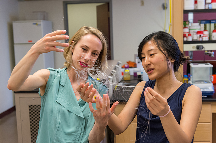 Researchers at MIT, under the direction of CSNE member and MIT professor, Polina Anikeeva, have been developing flexible neural implants for some time now; however, this research advance is significant because the stretchability of this new, rubbery, multifunctional fiber will better accommodate natural movement of the spinal cord in the body.
The spinal cord “undergoes stretches of about 12 percent during normal movement,” said Anikeeva in MIT News. “The fiber can stretch by at least 20 to 30 percent without changes in its properties.”
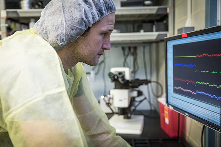
 The challenge of studying neurons in the spinal cord and the benefits of having a research tool such as this was underscored by the paper’s co-author, CSNE member and UW Postdoctoral Fellow in Neuroengineering, Thomas Richner, who conducted several of the lab experiments confirming the efficacy of these neural implants.
“It’s incredibly hard to create a neural interface with the spinal cord,” Richner said. “It’s much harder than making a neural interface with the brain or a peripheral nerve. The reason is that the spinal cord moves up and down within the vertebrae, and so it’s like trying to interface with a moving target.”
 Because this is the first device capable of providing both optical and electrical stimulation to neurons in the spinal cord, the new fiber shows promise to benefit not only future studies by CSNE members, but also spinal cord research conducted by members of the neuroscience and neural engineering communities at-large.
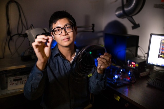
“We’re the first to develop something that enables simultaneous electrical recording and optical stimulation in the spinal cords of freely moving mice,” said the paper’s lead author, CSNE member and MIT graduate student, Chi (Alice) Lu in MIT News. “So, we hope our work opens up new avenues for neuroscience research.”
Research advance facilitated by the CSNE
The CSNE played a crucial role in this research advance, not only in terms of funding support, but also by laying the groundwork for quick and effective collaboration. The researchers at MIT had initially fabricated the neural implants and completed much of the experimental work necessary for their paper; however, one of the paper’s reviewers noted that more in vivo (in life) data was needed. The publication demanded a tight timeline, which could have proved problematic, but because of Anikeeva’s and Lu’s tight affiliation with the CSNE, they had already established working relationships with Richner and with Chet Moritz, CSNE faculty deputy director at the UW and Director of Moritz Lab. Richner and Moritz are experts in gathering and analyzing the type of in vivo data needed for this study, so getting the necessary expertise in a timely way was simply a matter of a quick email and a phone call.
 “Professor Anikeeva sent an email to Chet on Thursday night, saying ‘We need help getting more data to get through this review,’” Richner said, “and so, on Sunday I got on a plane to Boston and spent the following week helping them get whatever data needed to make sure it [the publication] got through review. We helped carry the baton through the last leg of the relay.”
As a result, a new and powerful research tool is on its way to being available to help better understand spinal cord injury, using the latest technologies available.
“There’s a growing set of genetic technologies that allow us to modulate neurons with light,” Richner said. “Professor Anikeeva is building a toolbox of micro-fabricated implantable hardware that meshes with these genetic technologies to enable researchers to reverse-engineer the spinal cord.”
For more information about this study, please contact Polina Anikeeva.