
 To better understand what is happening with neural signals in the brain, scientists continue to develop ways to simultaneously probe and manipulate neurons during simple tasks, like moving a cursor on a screen.
Now, a team of researchers at the Massachusetts Institute of Technology led by Polina Anikeeva, an assistant professor of materials science and engineering, have created polymer probes that will enable a more detailed manipulation and analysis of neural circuits deep in the brain than achievable before.
The findings were published January 19 in Nature Biotechnology. A study published in 2014 described the use of similar technology to record and stimulate neural activity in the spinal cord.
The MIT scientists used a thermal design process—which is commonly applied to glasses—to address some of the major limitations with other probes, including the formation of glial scars.
“We anticipated that our flexible devices would be more biocompatible than traditional probes, but we were somewhat shocked when we couldn't find them in the tissue,” Anikeeva said. “It was as if the brain has mistaken our probes for blood vessels.” The probes were studied in mice.
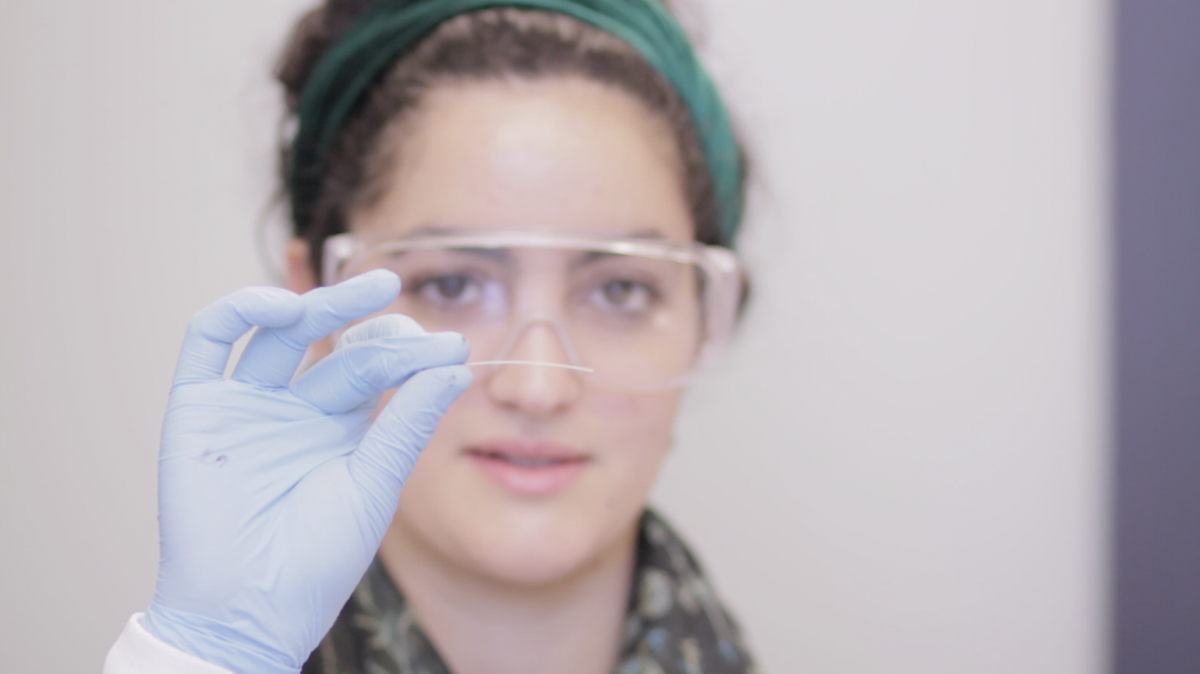
The polymer-based neural probes have feature sizes as small as five microns. An average human hair, in comparison, has a diameter of 100 microns.
According to the research team, which includes 10 other scientists, the probes formed stable brain-machine interfaces for at least two months. This, said Anikeeva, is significantly better than the current state-of-the-art in brain-machine interfaces.
“We recently took another look at some of our devices that remained implanted for approximately eight months, and we still find the same quality of recordings,” she said. “This was not possible previously with existing technologies.”
Anikeeva said she is hopeful that the new probes will become the foundation for brain and spinal cord interfaces for closed-loop co-adaptive bi-directional brain-computer interfaces that are at the core of the Center for Sensorimotor Neural Engineering.
“Being engineers at heart, we will continue pushing the resolution, multi-functionality, and biocompatibility of our devices by incorporating new polymers and composites,” she said, describing what’s next.
Anikeeva said that she and her team will collaborate with neuroscientists and physiologists on using the probes to map neural circuits and, in the future, as diagnostic tools. The team is working on future designs that incorporate smaller features and softer, more biocompatible polymers and potential surface modifications that aim to improve on the long-term compatibility and functionality of fiber-based multimodal neural interfaces.
Anikeeva is a principal investigator at the Center for Sensorimotor Neural Engineering. The study published in Nature Biotechnology was funded in part through the CSNE.