
When Aalap Dighe, PhD student in Mechanical Engineering at MIT, recently traveled to Seattle, he was thankful to escape the never-ending Boston snowstorms. But he wasn’t here just to escape the winter weather.
Dighe, who will graduate this summer, has traveled regularly to the University of Washington (UW) over the last few years as part of his role in the Center for Sensorimotor Neural Engineering (CSNE), an Engineering Research Center supported by the National Science Foundation.
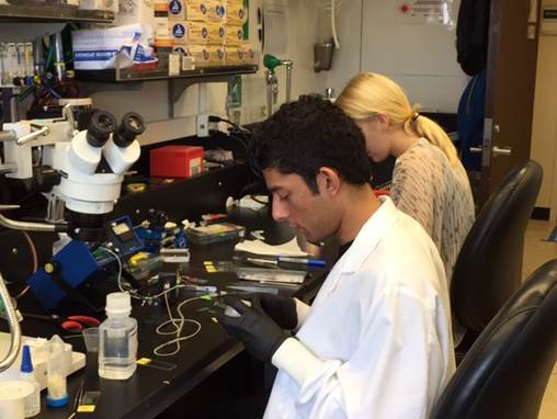 A member of MIT’s Biological Microtechnology and BioMEMS Group, Dighe has collaborated with Michael Sunshine, research scientist, and Aiva Ievins, PhD student in Neuroscience, in the Moritz Lab at the UW. Their goal? To build a better brain-computer interface, a device that can regularly record signals in the brain, which could one day help restore movement for people with motor disabilities.
“It’s been a joint effort,” Dighe said. Sunshine takes the lead on creating the printed circuit board, which supports and connects electronic components. Ievins evaluates tissue response and long-term longevity of the implants. “At the beginning of each design iteration, all three of us meet to discuss the different features of the device from a biological as well as from an engineering point of view, and how we can best optimize the various features, keeping the various trade-offs in mind,” he said.
On this recent trip, Dighe and the UW team were testing out a third-generation device currently known as the MIT Probe. This naming convention is similar to other devices on the market, including the Utah Array—created at the University of Utah—and the Michigan Probe—created at the University of Michigan.
Two features of the device make this probe unique. For starters, the tips of the device are really small, under 10 to 20 microns. (One micron = 1/25th of a thousandth of an inch.)
Device will record from brain cells
“The goal of the device is to record electrical activity from small group of cells and, ideally, single cells in the brain for really long periods of time, months and years, eventually,” said Dighe. “If you can do that, you can enable closed-loop devices that could restore movement for people with paralysis. The reason that you don’t have these devices right now is because you don’t have interfaces with electrodes that can reliably record for that long.”
 When existing brain-computer interfaces are implanted, the device stops working after a few weeks or months due to a sheath of glial cells (the immune cells in the brain) that forms around the device, electrically insulating it from the neurons from which it is trying to record. This glial sheath is the body’s way of protecting itself from a foreign object in the brain.
The body reacts to any foreign body; think of the response to getting a splinter, for example. Your skin turns red, revealing the signs of infection. Your body will eventually push the splinter out of the body in some cases, too, if you can’t remove it with tweezers.
“That becomes a problem when you’re trying to record brain signals,” said Dighe. “Once the glial sheath forms around an electrode, the signal drops to noise levels. There is no way that you can ‘listen harder’ from brain cells once a glial sheath forms.”
New device targets the glial sheath
The MIT Probe is also equipped to pierce through the glial sheath once it forms. According to Dighe, piercing through the glial sheath is a fairly common technique performed with microwires. “Researchers use microwires to pierce the glial sheath and temporarily recover the signals that have been lost. They currently do it with relatively big wires, and so the glial sheath eventually forms again. You recover signals, but they die down again.”
The UW-MIT team is combining these two approaches—the tiny tips and the glial sheath-piercing process. Initial tests show that the mechanical and electrical features of the device work. They plan to implant the device, wait for the tissue reaction to happen—which takes a few weeks —and then poke through the sheath with the tiny tips. They are currently testing the device with animal models.
If all goes well, the MIT Probe could one day be an alternative to existing products on the market like the Utah Array.
Ian Dryg, graduate student in the UW Dept. of Bioengineering and CSNE member, is also collaborating on this research. Dryg works in the lab of Bill Shain, UW professor of Neurological Surgery. Shain, also a CSNE member, is considered a leading expert on glial reaction. His lab is based at Seattle Children’s Research Institute.